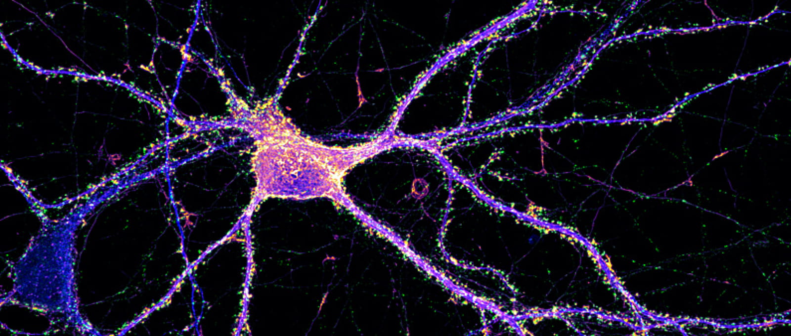
Neurone d'hippocampe étiqueté pour l'actine (orange), la synapsine (vert) et map2 (bleu).
AMU/CNRS Christophe Leterrier

Institute of NeuroPhysiopathology

The Institute of NeuroPhysiopathology (INP, UMR7051), directed by Michel Khrestchatisky, was created on January 1, 2018, under the supervision of the CNRS and AMU and will be housed in renovated premises within the Faculty of Medicine of Marseille. This operation is part of the NeuroTimone project aimed at forming a pole of excellence in Neurosciences. The INP has the particularity of combining public and private research within a Joint Research Laboratory, notably thanks to the partnership with the biotechnology company VECT-HORUS specialising in the brain targeting of therapeutic agents. Technology platforms (PFRN, PFNT) in the fields of imaging, molecular interactions and stem cells (iPS) are labelled and backed by the INP, thus strengthening its research potential. The INP brings together nearly 150 people divided into 11 research teams. With complementary skills in animal and in vitro models, the laboratory teams study cellular and molecular mechanisms that govern the neural bases of brain function and dysfunction. Several teams aim to better understand glioma biology and identify new therapeutic targets. Others focus on neuronal plasticity and memory processes, as well as neurodegenerative and neuro-inflammatory processes involved in developmental disorders, Alzheimer’s disease, multiple sclerosis and spinal cord injuries. The teams enhance their research by developing: i) molecular therapy approaches, with agents that control the action of matrix proteases, neuro-inflammation, leukocyte recruitment, axonal regeneration, tumour progression; ii) cell therapy approaches in the central nervous system, with nasal olfactory mucosal stem cells and neural cells derived from induced pluripotent cells (iPS cells); iii) innovative vectorization strategies for addressing drugs or imaging agents to the central nervous system across the blood-brain barrier.

Pictures from the INP laboratory

Research Teams

11 teams the INP are studying on a fundamental level, through animal and cell models, at different levels of integration, and in various pathological contexts, the interactions between neuronal, glial, endothelial and immune cells, and their roles in the plasticity of neural networks, in biological clocks and in cognitive function. The teams of the Institute study during development and aging different conditions including Alzheimer’s disease, multiple sclerosis, neuroinflammatory and neurodegenerative processes, glioblastoma, tumorigenesis, angiogenesis and alterations of the barrier blood-brain.

Discover the NeuroTimone stem cell platform at the INP (don't forget to enable the English subtitles)

BBB and neuroinflammation

Description

The central nervous system (CNS) plays a major role in the functioning of the body and therefore benefits from a vascular system particularly adapted to its protection, the blood-brain barrier (BBB). It effectively protects the CNS from the entry of potentially neurotoxic endogenous or exogenous agents, but considerably limits the passage of pharmacological, therapeutic or imaging agents. The BBB thus appears to be a major obstacle to the treatment of CNS pathologies while the prevalence of CNS pathologies is increasing and the therapeutic area of the CNS is one of the main pharmaceutical markets. The molecular and cellular mechanisms that regulate the weakening and permeability of the BBB in a neuro-inflammatory context are still poorly understood and are central to our projects.

In addition, we are interested in receptor families involved in transport processes across the BBB and are developing vector molecules that specifically bind to these receptors to promote the transport of therapeutic and imaging agents across this barrier. These projects are carried out in partnership with the company Vect-Horus, spin-off of the laboratory.


Team leader

Michel Khrestchatisky  Total : 3 HDRs.

Techniques

  • Molecular biology
  • Biochemistry
  • Cell culture
  • Immunostaining, histology, flow cytometry
  • Microscopy
  • Animal surgery, stereotaxy
  • Pharmacology
  • Animal behavior
  • Brain imaging – Animal
  • Medical data analysis
  • Bioinformatics

Research axes

  1. Neuromodulation of BBB permeability
  2. Vectorization of molecules through the BBB.

Keywords

Blood-brain barrier, neuro-inflammation, endothelial cells, transcriptional analyses, proteomics, therapeutic agents, imaging agents, vectorization, Alzheimer’s, glioblastoma.

Thematics
  • Animal Cognition And Behavior
  • Disorders Of The Nervous System
  • Novel Methods And Technology Development

Neural Degeneration and Plasticity

Description

Understanding and fighting Alzheimer’s disease is one of the most challenging endeavors of modern neuroscience. Alzheimer’s disease is the most devastating neurodegenerative disorder with a major socio-economic burden. Despite a variety of therapies currently investigated, the discovery of a cure does not seem to be within reach at this point. Therefore, there is an urge in identifying new targets in the triggering/progression of the disease and the underlying molecular mechanisms.

Our global objective is to better understand the role of some proteolytic pathways at the crossroads of three major pathogenic processes: neuroinflammation, amyloidogenesis and synaptic dysfunctions. Our original findings place membrane bound matrix metalloproteinases (MT-MMPs) as new actors and potential new therapeutic targets in Alzheimer’s pathogenesis because they promote neuroinflammation and amyloidogenesis, and alter synaptic transmission.

Our research is therefore supported by two main objectives: i) increase our understanding of the pathophysiological mechanisms of Alzheimer’s disease, and ii) develop and validate innovative therapeutic strategies on the basis of newly discovered targets. We use state of the art techniques of molecular and cellular biology, biochemistry, cell and tissue imaging, pharmacology, electrophysiology and animal behavior, on in vivo and in cellulo models of the pathology, in collaboration with INP teams and worldwide.

Team leader

Santiago Rivera  Total : 2 HDRs.

Techniques

  • Molecular biology
  • Biochemistry
  • Cell culture
  • Immunostaining, histology, flow cytometry Microscopy
  • Calcium imaging
  • Electrophysiology (on slices or cells)
  • Animal surgery, stereotaxy
  • Pharmacology
  • Animal behavior
  • Medical data analysis

Research axes

  1. Alzheimer’s disease
  2. Pathophysiological mecanisms and novel therapeutic strategies

Keywords

Alzheimer’s disease, neurodegeneration, neuroinflammation, proteolysis, metalloproteinases, therapeutic strategies, iPS cells, Crispr/Cas9, transgenic mouse, whole cell patch clamp.

Thematics
  • Animal Cognition And Behavior
  • Disorders Of The Nervous System
  • Excitability, Synaptic Transmission, Network Functions
  • Novel Methods And Technology Development

NOSE. Nasal Olfactory Stemness and Epigenesis

Description

Our team co-discovered and characterised the adult human and rodent nasal olfactory stem cells. We use these unique cells to investigate the molecular mechanisms underlying the symptoms of Autism Spectrum Disorders. An abnormal expression of an enzyme (MOCOS) was unveiled in patients’ stem cells. We now assess the role of this enzyme during brain development and try to identify drugs that restore a physiological expression of MOCOS.

The team also uses human olfactory stem cells for repairing the pathological brain. We performed studies based on the transplantation of olfactory stem cells in models of amnesia and Alzheimer’s disease. We are currently devising new protocols for differentiating stem cells into dopaminergic neurons. The team is engaged in translating our basic research into clinical applications. Trials dedicated to nerve and bone repair are in preparation.

In parallel, in order to improve functional recovery, the team assesses the immuno-modulatory and neurotrophic roles of vitamin D. Preclinical studies on animal models of nerve section, paraplegia and Alzheimer’s disease are encouraging.

Team leader

François Féron. Total : 5 HDRs.

Techniques


  • Molecular biology
  • Biochemistry
  • Cell culture
  • Immunostaining, histology, flow cytometry
  • Microscopy
  • Animal surgery, stereotaxy
  • Pharmacology
  • Animal behavior
  • Brain imaging – Animal
  • Medical data analysis
  • Bioinformatics
Research axes

  1. Olfactory stem cells, a diagnosis tool
  2. Olfactory stem cells for cell therapy
  3. Vitamin D, a neurosteroid hormone

Keywords

Stem cells, plasticity, cell therapy, vitamin D, autism, Alzheimer’s disease, circadian rhythms, non-coding RNA, Prader-Willi syndrome, olfactory receptors

Thematics
  • Animal Cognition And Behavior
  • Development Of The Nervous System 
  • Disorders Of The Nervous System
  • Excitability, Synaptic Transmission, Network Functions
  • Human Cognition And Behavior
  • Sensory Systems

Genes, Rhythm and Neurophysiopathology

Description

The daily change of day and night due to the Earth's rotation along its axis, has led all living organisms on this planet to develop internal clocks called circadian clocks that allow them to adapt and anticipate this daily change in the environment.

The team's research focuses on the molecular mechanisms of these circadian clocks, particularly those involving long non-coding RNAs.

Knowing that profound alterations in our biological clock can be observed in patients suffering from pathologies of the central nervous system, the team's mission is also to study the role played by genes, proteins and long non-coding RNAs involved in the functioning of the circadian clock, in brain diseases, in particular neurodevelopmental diseases such as Prader-Willi syndrome.

Team leader Techniques

  • Molecular Biology
  • Biochemistry
  • Cell culture
  • Immunostaining, Hybridization in situ
  • Microscopy
  • Pharmacology
  • Animal behaviour
  • Analysis of NGS data
  • Bioinformatics

Research axes

  1. Characterization of the molecular mechanisms involved in the control of circadian gene expression, in particular those involving ncRNAs such as Neat1 or Malat1
  2. Characterization of circadian anomalies associated with the physiopathology of Prader-Willi syndrome and identification of the genes involved in these dysfunctions

Keywords

Circadian rhythms, gene expression, post-transcriptional regulation, non-coding RNA, paraspeckles, Prader-Willi syndrome, NGS bioinformatics analyses

Thematics
  • Development Of The Nervous System 
  • Disorders Of The Nervous System
  • Excitability, Synaptic Transmission, Network Functions
  • Human Cognition And Behavior
  • Sensory Systems

Neurobiology of mnesic processes

Description

The team is working on changes in the neurobiological substrate during learning and memory processes, including in pathological conditions. The underlying hypothesis is that durable memory traces proceed from the marking of specific neural networks. These synaptic plasticity mechanisms result from molecular and structural changes that largely remain to be identified and integrated at the behavioral level.

In mice, we developed a new olfactory test, “the tubular olfactory maze” where we test the effect of molecules or genetic manipulations on different memory subcategories. We observed specific structural changes in the dorsal and apical dendrite density of CA1 pyramidal cells of the hippocampus, specifically amplified by a 5-HT4 receptor agonist known for his promnesic effect. We are currently using this test in a model of Alzheimer’s disease (5XFAD transgenic mice).

With the same aim of creating animal models to study memory, we developed a deferred task paradigm in mice that highlights specific deficits as observed in patients with frontal lobe syndrome, including a perseveration behavior.

Finally, with a view to overcome the memory deficits in our amnesic syndrome models in mice, we performed intra-hippocampal transplantation of adult stem cells from the human olfactory mucosa. Following these grafts, we observed a recovery of memory capacity and the ability of these cells to acquire a neural or glial phenotype. We are currently conducting autologous grafts in rat models of amnesia and spinal cord injuries.

Team leader

François Roman. Total : 3 HDRs.

Techniques

  • Molecular biology
  • Biochemistry
  • Cell culture
  • Immunostaining, histology, flow cytometry
  • Microscopy
  • Electrophysiology (in vivo)
  • Animal surgery, stereotaxy
  • Pharmacology
  • Animal behavior
  • Psychophysical tests
  • Brain imaging – Animal

Research axes

  1. Effect of an agonist of 5-HT4 receptors on the dendrite structure of hippocampal neurons
  2. Memory performance in a model of Alzheimer’s disease (5XFAD transgenic mice) in a tubular olfactory labyrinth
  3. Deferred task tests in a mouse model of frontal syndrome
  4. Autologous transplants of adult stem cells in rats with amnesia or spinal cord injuries.

Keywords

Synaptic plasticity, learning, memory, 5HT4 receptors, mnemonic disorders, transplantation, olfactory stem cells, Alzheimer’s disease, frontal disorders, spinal cord injuries

Thematics
  • Animal Cognition And Behavior
  • Disorders Of The Nervous System
  • Excitability, Synaptic Transmission, Network Functions
  • Human Cognition And Behavior
  • Sensory Systems

Neuro-inflammation and multiple sclerosis

Description

The « Neuroinflammation and Multiple sclerosis » team is an emerging research team issued from the « BBB and Neuroinflammation » team. It is coordinated by Sophie Desplat-Jégo, MCU-PH, MD, PhD , specialized in medical immunology. SDJ has developed a research topic on the role of the cytokine TWEAK, member of the TNF family during neuroinflammation especially in the pathological context of multiple sclerosis (MS) and her group has generated reference articles that describe the deleterious effects of TWEAK in the CNS during MS and its animal model, EAE. In the last years, the TWEAK project has focused on the study of biological effects of TWEAK on the properties of the BBB. In the coming years, the team will pursue studies on neuroinflammation and involvement of TWEAK in MS.

Research axes

  1. To further evaluate the relevance of targeting TWEAK/Fn14 pathway in CNS for treating MS
  2. Characterize biological consequences of upregulation of soluble TWEAK and TWEAK-binding antibodies production during EAE /MS
  3. To determine if TWEAK is subject to polymorphism and to determine functional consequences of this polymorphism

Thematics
  • Disorders Of The Nervous System

Stem Cells, Disease Modeling and Neuroregeneration

Description

Our research team is studying the molecular and cellular mechanisms driving pathogenic processes in the human brain, with specific focus on two diseases for which the lack of treatments urge the need for new insights: 1) the Alzheimer’s disease (AD); and 2) brain tumours, namely gliomas.

Permanent questioning is the seed of Science. As such, our research group is wondering: How genetic risk factors impact on AD pathogenesis? Is neuroinflammation one of the driving forces in AD pathology? Is the astrocyte a pivotal cellular determinant in AD? Noteworthy, we are also working on the validation of new therapeutic targets in AD. Besides, we are also developing human cell-based models aiming to recapitulate gliomagenic processes, allowing the study of the mechanisms at stake and offering the possibility to screen/test therapeutic molecules against brain cancer cells.

To quench our thirst for new knowledge, our team is leveraging on innovative and cutting-edge strategies. The core of our research relies on the development/use of cell reprogramming-based disease modelling strategies (human induced pluripotent stem cells especially) as well as genome editing technologies (CRISPR-Cas9-based); which we combine with a battery of techniques (molecular and cellular biology, biochemistry, cell imaging, pharmacology) to address our questions.

Team leader

Emmanuel Nivet  Total : 1 HDR 

Techniques utilisées

  • Molecular biology
  • Biochemistry
  • Cell culture
  • Immunostaining, histology, flow cytometry
  • Microscopy
  • Calcium imaging
  • Electrophysiology
  • Pharmacology

Research axes

  1. Alzheimer’s disease
  2. Gliomagenesis
  3. Cell reprogramming

Keywords

Alzheimer’s disease, glioma, cell reprogramming, neuroinflammation, CRISPR-Case9, genetic risk factors.

Thematics
  • Disorders Of The Nervous System
  • Novel Methods And Technology Development

NeuroCyto: the neuronal cytoskeleton in physiology and disease

Description

“NeuroCyto: the neuronal cytoskeleton in physiology and disease” is a lab that started in 2017 and is part of the Neuropathophysiology Institute (INP, CNRS-Aix Marseille University UMR 7051) in beautiful Marseille, France.

The team currently has six members, and we welcome trainees all year long. Motivated students are always welcome to contact us! We aim at building a thriving team to make the best possible science by nurturing openness, exchange, and the excitement of discoveries big and small.

At NeuroCyto, we want to understand how neurons are organized at the cellular level. How do they differentiate, then build and maintain their complex arborization? How do they establish and conserve their polarity, with the axon and dendrites allowing to send and receive signals? Numerous processes contribute to this organization: elaboration of the cell architecture (thanks to the cytoskeleton), protein transport inside the cell (with diffusion and motor proteins), segregation into distinct compartments (such as axon, synapses, dendritic spines…). The NeuroCyto team applies advanced microscopy techniques to directly observe molecular assemblies at the nanoscale in neurons, revealing how they organize the neuron and shape its physiology.

Team leader

Christophe Leterrier. Total : 1 HDR.

Techniques

  • Molecular biology (PCR…)
  • Biochemistry (Western blot…)
  • Cell culture
  • Immunostaining, histology, flow cytometry
  • Microscopy (fluorescence, confocal, electronic…)

Research axes

  1. Cell biology of the neuron
  2. Axonal cytoskeleton
  3. Super-resolution microscopy

Keywords

Neuronal cytoskeleton, axonal, super-resolution microscopy, neuron organization.

Thematics
  • Development Of The Nervous System 
  • Excitability, Synaptic Transmission, Network Functions
  • Novel Methods And Technology Development

Gliomagenesis and microenvironment

Description

Our GlioME team develops projects to better understand human glioma biology, to identify new targets, and to evaluate promising anticancer therapies using appropriate cellular and pre-clinical models.

Gliomas are the most common intracranial tumors in humans. The most malignant is glioblastoma (GBM, grade IV), with an incidence of 3–5 out of 100,000 persons in Western countries. GBs are lethal tumors and even optimal surgical resection, followed by chemotherapy and irradiation, results in a median survival of about 12–15 months. We trust that a comprehensive understanding of glioma cell-of origin as well as the intricate micro-environmental landscape of these tumors will result in the development of novel therapeutic strategies.

Therefore, our team focused on:

1. Setting up performing preclinical glioma models and identify new targets

2. Identify glioma biomarkers and participating to clinical trials

We are also involved in basic science consortia dedicated to nanoparticles design and development for brain tumor treatment.

Team leader Techniques

  • Molecular biology
  • Biochemistry
  • Cell culture
  • Immunostaining, histology, flow cytometry
  • Microscopy
  • Pharmacology
  • Brain imaging – Animal
  • Medical data analysis
  • Bioinformatics

Research axes

Fondamental

  1. Deciphering the role of A2B5, a cancer stem cell marker, in gliomagenesis
  2. Deciphering the role of the Inhibitors of Apoptosis (IAPs) in the regulation of GB progression, microenvironment (angiogenesis, hypoxia and inflammation) and stemness
  3. Deciphering the role of Microtubule Dynamics (EB family) in the regulation of GB growth (microenvironment and stemness)

Translationnels : Biomarkers / Therapy

  1. Identifying biomarkers for diagnosis, prognosis, response to treatment in gliomas and glioneuronal tumors
  2. Deciphering the biological role of MMP2 and MMP9 in GB development and resistance to anti-angiogenic therapies
  3. Improving nanoparticle (NPs) delivery in the brain for theranostic applications
Keywords

Human gliomas, microenvironment, stem cells, A2B5, IAPs, microtubule dynamics, nanoparticles, diagnostic/prognostic biomarkers, predictive markers of response to treatment, therapeutic targets, preclinical models.

Thematics
  • Disorders Of The Nervous System
  • Novel Methods And Technology Development

Cytoskeleton and Neurophysiopathology

Description

We aim to identify biomarkers and new pharmacological compounds active for cancer and neurodegenerative diseases with specific focus on tubulin cytoskeleton and Tau protein, Rho-GTPases, integrins, aggregative proteins and redox signaling

Team leaders

KOVACIC HervéPEYROT Vincent, Total : 5 HDRs.

Techniques

  • Molecular biology
  • Biochemistry
  • Cell culture
  • Immunostaining, histology, flow cytometry
  • Microscopy
  • Pharmacology
  • Bioinformatics
  • Biophysic of molecular interaction

Research axes

  1. Tau post-translational modification (PTM): from molecule to bedside
  2. Identification of biomarkers in neurology and neuro-oncology
  3. NADPH oxidase as mediator of neurotoxicity

Keywords

Microtubules, actin, Tau, proteinopathies, NAPDH oxidases, glioblastoma, resistance to chemotherapy, new therapeutic agents.

Thematics
  • Disorders Of The Nervous System
  • Novel Methods And Technology Development

Oligodendroglial Cell Lineage in Aging and in Disease

Description

In the central nervous system, oligodendroglial cells represent an abundant cell population with critical functions, as they sense, regulate, and provide insulation and trophic support to neurons. From oligodendrocyte progenitor cells to myelinating oligodendrocytes, the oligodendroglial lineage is therefore crucial for normal neurological functions, such as brain plasticity. With age, in parallel with a profound decline of cognitive abilities, oligodendroglial capacities have been shown to decline. Defective myelination has been attributed to age-related epigenomic, transcriptomic and phenotypic changes in adult oligodendroglial cells, affecting their myelinating properties.

Our team addresses: How aging affects oligodendroglial cells in the central nervous system? How can it directly impact brain cognition in aging and in neurodegenerative diseases? Can we rejuvenate old oligodendroglial cells?

By challenging our aging-like oligodendroglial-specific mouse model and age-related disease models with behavioral tests, we address the role of oligodendroglial cells in aging and in psychiatric and neurodegenerative diseases. We use transcriptomic approaches and in vitro assays to identify and characterize the molecular mechanisms that regulate neuro-oligodendroglial interaction, from a specific oligodendroglial point of view. We are also leveraging epigenetic activators, in combination with non-invasive delivery tools, to reactivate cells in our aging and disease models.

Team leaders

Moyon Sarah

Techniques

  • Molecular biology (PCR...)
  •  Biochemistry (Western blot...)
  •  Cell culture
  • Immunostaining, histology, flow cytometry
  • Microscopy (fluorescence, confocal, electronic...)
  • Animal surgery, stereotaxy
  • Bioinformatics

Research axes

1. Aging oligodendroglial cells and cognition

2. Oligodendroglial cells in neurodegenerative diseases (Alzheimer’s Disease)

3. Neuro-oligodendroglial cell communication

4. Epigenetics approaches to rejuvenate the brain

Keywords

Oligodendrocytes, Myelination, Neuro-oligodendroglial communication, Aging, Cognition, Neurodegeneration, Alzheimer’s Disease, Epigenetics, TET1-mediated activation

Thematics
  • Animal Cognition And Behavior
  • Disorders Of The Nervous System
  • Novel Methods And Technology Development

Angiogenesis and tumor microenvironment

Description

Glioblastoma (GBM) is a devastating brain tumour characterized by local invasion, microvascular proliferation, and therapeutic resistance. The highly infiltrative nature of glioma cells makes complete surgical resection unlikely, and 90% of tumors recur. High-grade gliomas are among the most angiogenic of all tumors. Adrenomedullin (AM) is a 52 aminoacid C-terminal amidated peptide widely expressed in a variety of tumor types and was shown to be mitogenic for many human cancer cell lines in vitro. AM binds to and mediates its activity through the G protein-coupled receptor calcitonin receptor-like receptor (CLR), with specificity for AM being conferred by the receptor activity modifying protein -2 (RAMP2) and -3 (RAMP3).

The genetic ablation of AdM, calcrl, Ramp2 or the enzyme responsible for functional AM amidation, peptidylglycine α-amidating monooxygenase (PAM) all result in midgestational lethality associated with severe interstitial edema and cardiovascular defects. In vivo studies highlight the significance of AM as an important factor to promote tumor growth and to affect the tumor microenvironment by inducing pathologic angiogenesis and lymphangiogenesis. Accumulating studies suggest a new role for AM as a cross-talk molecule that integrates tumor and microenvironment stroma cells underlying promotion mechanisms to facilitate angiogenesis and tumor growth.

Several in vivo studies have shown a regression of tumor neovessels and growth upon the treatment with neutralizing AM antibodies, AM receptor antagonist, or AM receptor interference. These findings highlight the implication of AM in the progression of tumor growth and angiogenesis, suggesting that targeting the AM system may be a useful therapeutic strategy in brain cancer. Therefore, understanding the molecular mechanisms by which AM can determine the integrity of tumor neovessels will be the main focus of our research with the use of GBM as model to pursue our projects.

Team leader
Research axes

  1. Role of AM in the recruitment of pro-vascular and pro-angiogenic bone marrow-derived cells in tumor neovascularization
  2. Role and mechanisms of action of AM in the tumor blood vessels stabilization in GBM
  3. Role of AM in the neoangiogenesis and/or vasculogenesis during glioma recurrence after radiotherapy
  4. Expression of the AM system in glioblastoma-associated vascular cells
  5. Therapeutic issues: inhibition of glioblastoma tumor growth by targeting only AM system using monoclonal antibodies or in combination with classical or innovative drugs

Vect-Horus

Description

VECT-HORUS is a biotechnology company that designs and develops peptide-based vectors that facilitate the delivery of drugs or imaging agents into organs, notably into the brain, and to tumors. The vectors target receptors involved in “Receptor Mediated Transport” (RMT, a physiological system for the transport into cells of endogenous substances). By combining drugs or imaging agents to its vectors, VECT-HORUS allows them to cross biological barriers that restrict access to their target, notably the blood-brain barrier (BBB).

For more information, visit their website.


See more
Your opinion is important to us

Description de la soumission d'un avis
Your vote :
Your opinion is important to us
Publications