Description de la soumission d'un avis
Your vote :

The Center for Magnetic Resonance (MR) in Biology and Medicine (CRMBM) conducts translational research by developing and applying MR methods and instruments (RM) to explore the morphology, metabolism and physiology of human diseases and associated animal models (rodents). With the support of methodological and engineering teams, our research teams aim at (i) better characterizing healthy and pathological states of the central nervous, cardiovascular and musculoskeletal systems and (ii) defining new diagnostic and/or therapeutic strategies.
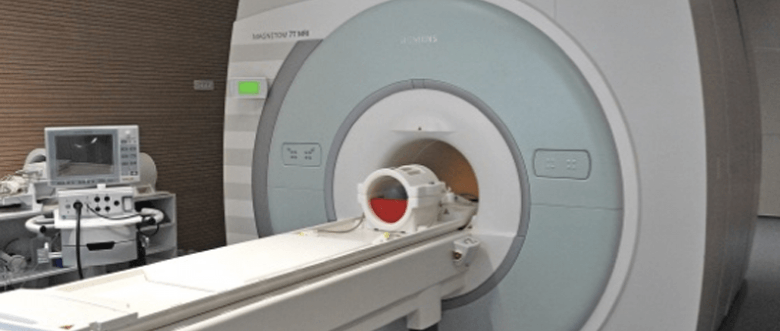
Research is conducted on state-of-the-art MR scanners for rodents at CRMBM (Horizontal 4.7T, vertical 11.75T, and horizontal 7T Bruker systems) and for humans at CEMEREM, the clinical site of CRMBM (Timone University Hospital), equipped with 3 clinical Siemens MR scanners (1.5T, 3T and the first 7T whole-body MR scanner in France).
The CRMBM team, Professor Ranjeva’s team, located at the CEMEREM and working on the human central nervous system, is affiliated to the neuroscience master’s program and can thus train neuroscience master’s students.
The CNS team aims at providing relevant innovative in vivo and non-invasive MRI parameters on patients and rodent models for the better understanding of the pathophysiology, structural abnormalities and dysfunction accompanying or causing some of the main neurological diseases such as multiple sclerosis, epilepsy, and spinal cord injury impacting public health, in close relationships with clinicians from the team and external collaborators specialized also in Alzheimer’s disease, schizophrenia and brain maturation.
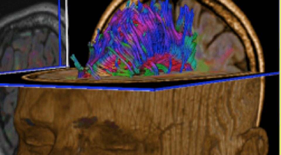
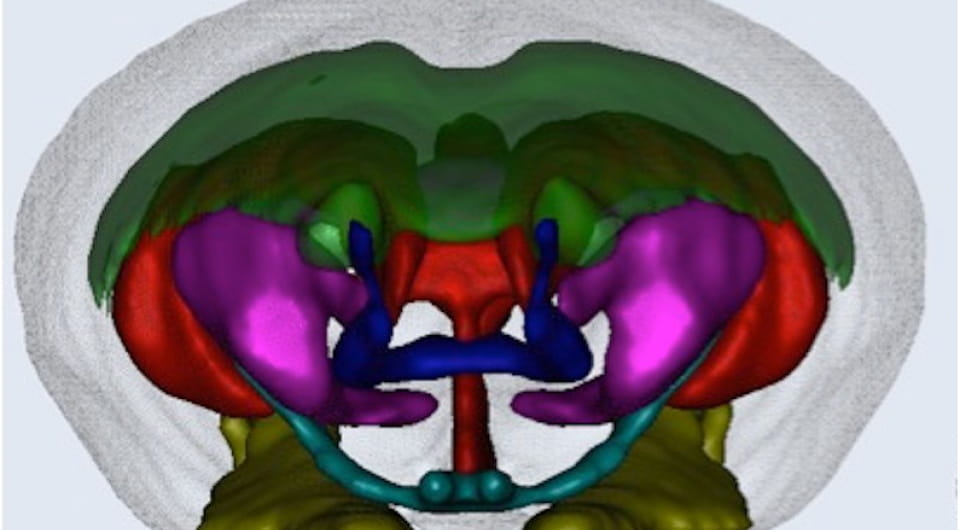
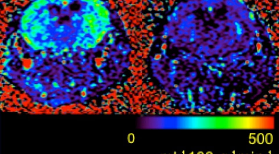
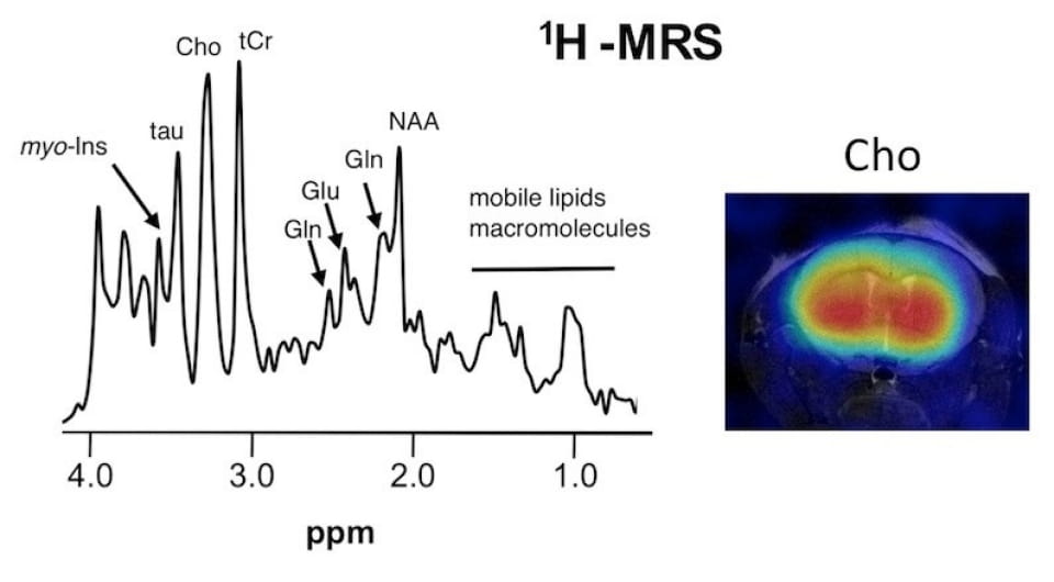
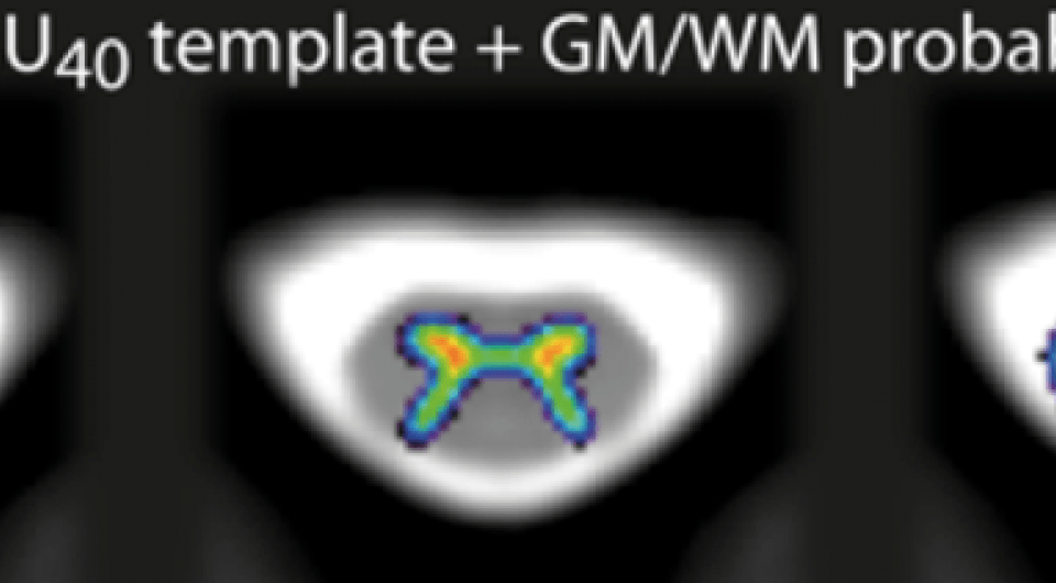
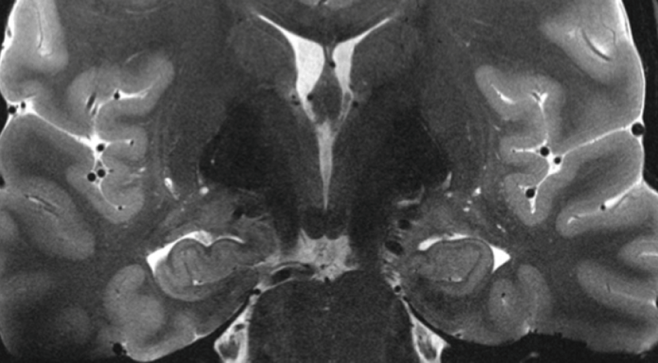
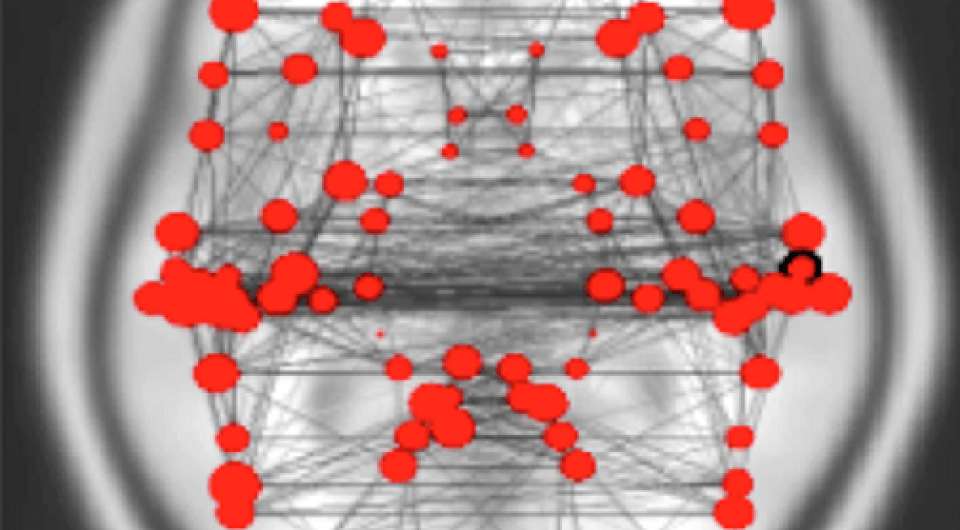
MRI explorations are conducted on high field (1.5T, 3T) and ultra high field (7T) clinical MR scanners and on preclinical MR scanners (4.7T, 7T, 11.75T) using advanced MR methods (Diffusion MRI, fMRI, rs-fMRI, MRSI, X-nucleus MRI, QSM, ASL, MTR, …).
Jean-Philippe Ranjeva, Angèle Viola Total : 9 HDRs.
Characterization of the pathophysiology and clinical consequences of central nervous system pathologies using advanced MRI techniques and neuropsychological and clinical data collected from patients with MS, epilepsy, amyotrophic lateral sclerosis, Alzheimer’s disease, spinal cord injury, schizophrenia and Parkinson’s disease.
Advanced Magnetic Resonance Imaging, fMRI, Spectroscopic MRI, Brain, Spinal Cord, Multiple Sclerosis, Epilepsy, Alzheimer, Amyotrophic Lateral Sclerosis, Stroke, Parkinson, High Field and Ultra High Field clinical and preclinical MR scanners.