
Description de la soumission d'un avis
Your vote :
The Institut de Neurosciences de la Timone (INT, UMR7289) is a prominent research institute within Aix-Marseille University and CNRS, located in the Timone campus (Marseille). Created in 2012, INT was founded with the core idea to cover a few Central Nervous System (CNS) functions, now organized in two thematic axes: dynamic sensory and motor functions, and adaptive responses to environmental changes. INT is rich and diverse with 13 dynamic teams covering different approaches (neurobiological, integrative & cognitive neuroscience), using a variety of experimental models (rodents, non-human primates & human subjects), in both healthy and pathological conditions.
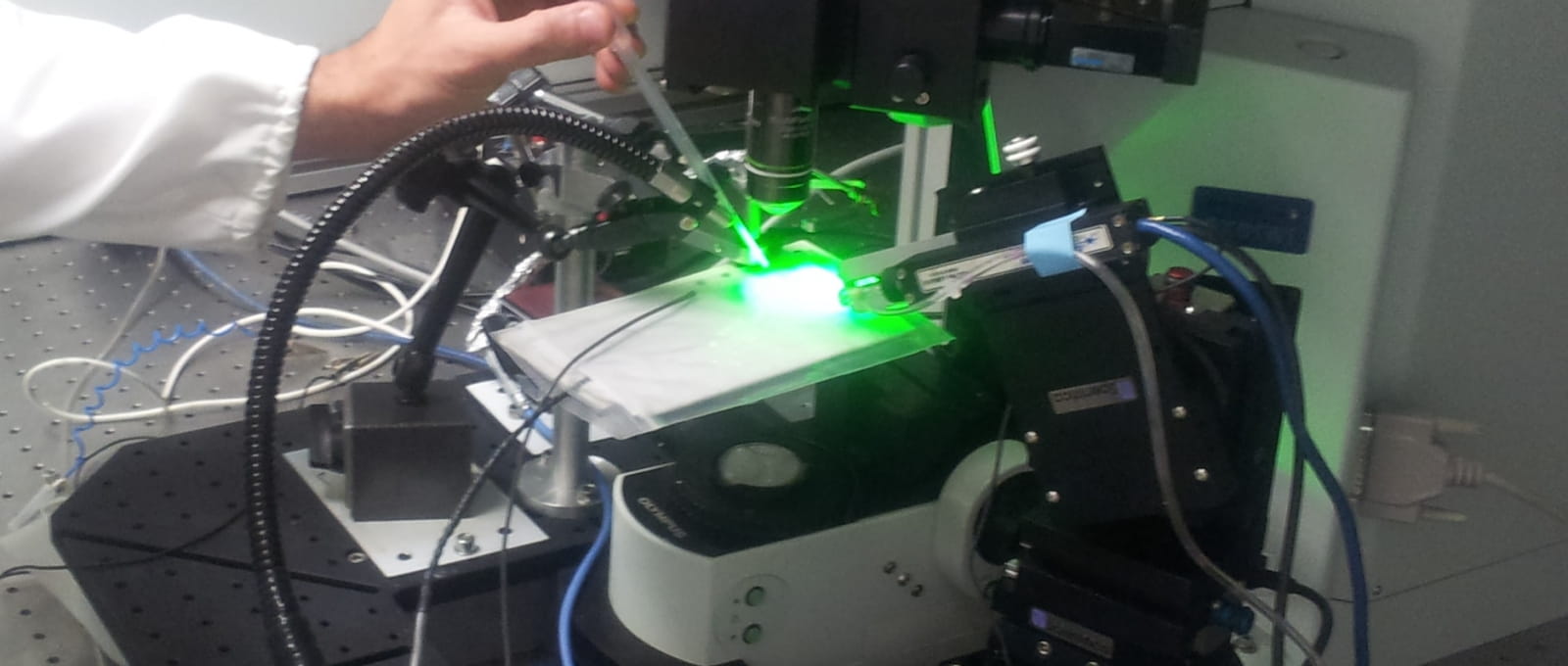
This exploration extends across three main structures of the central nervous system: the spinal, cortical & basal ganglia networks. One of the main objectives of INT is now to launch ambitious collaborative projects to study CNS functions across scales, structures, approaches and models. To reach this goal, INT relies on state-of-the-art technology grounded in theoretical principles, facilitated by its interdisciplinary centers: The NeuroTechCenter to develop and use advancing electrophysiological or photonics techniques, and the CoNeCT center to anchor our projects in computational and theoretical neurosciences. Moreover, INT benefits from well-structured facilities , including: support teams for administration and computer resources, sharing technical expertise in neurobiology, neurosurgery and instrumentation, and functional exploration of the nervous system, such as MRI and photonics.

Discover INT’s 13 interdisciplinary teams working on the multi-scale dynamics of neural networks in a functional context and the development of the brain and cognition.
Frédéric Brocard
Find out more, click on the following link: here
Rod O'Connor
Find out more, click on the following link: here
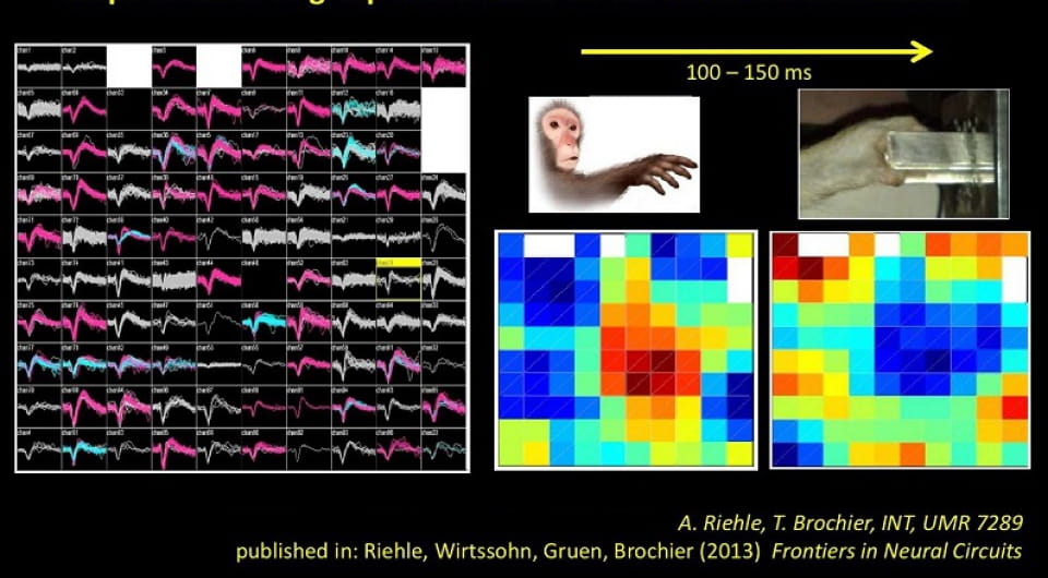
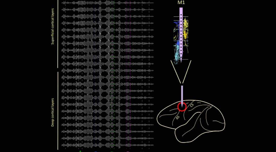
Bjørg Kilavik & Thomas Brochier
Find out more, click on the following link: here
Sophie Denève & Frédéric Chavane
Find out more, click on the following link: here
Guillaume Masson
Find out more, click on the following link: here
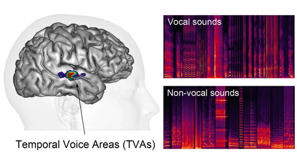
Pascal Belin
Find out more, click on the following link: here
Christelle Baunez
Find out more, click on the following link: here
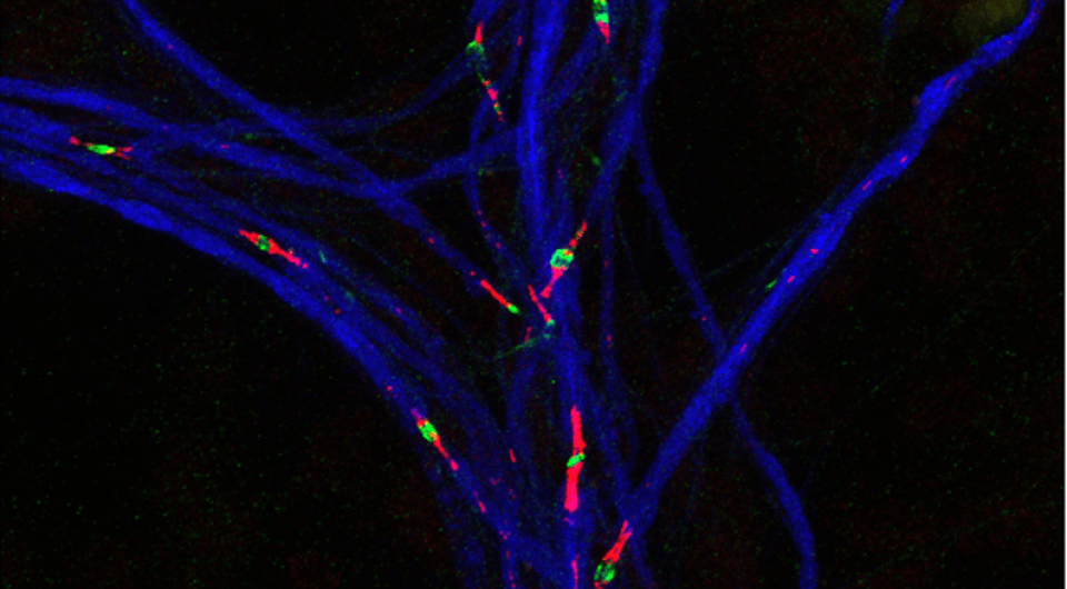
Eduardo Gascon
Find out more, click on the following link: here
Christine Deruelle
Find out more, click on the following link: here
Olivier Coulon
Find out more, click on the following link: here
Nicolas Wanaverebecq
Find out more, click on the following link: here
Jean-Marc Goaillard
Find out more, click on the following link: here
Andrea Brovelli