Description de la soumission d'un avis

Mediterranean Neurobiology Institute

- A developmental scaffold for the organization of adult cortical networks
- Neuronal coding of space and memory
- Early activity in the developing brain
- Adolescence and developmental vulnerability to neuropsychiatric diseases
- The neural bases of sensorimotor learning
- NICE2: Neonatal, Infantile and Childhood Epilepsies and Encephalopathies
- Neuronal coding and plasticity in epilepsy
- Molecular basis and pathophysiology of cortical development disorders
- Early life imprinting and neurodevelopmental disorders
The Institute of Mediterranean Neurobiology (INMED) is a leading Neuroscience Centre affiliated to INSERM and Aix-Marseille University. INMED investigates the development and plasticity of neuronal synapses and circuits in health and disease. Recently, the center became a pioneering leader in the field of Developmental Systems Neuroscience.
Over the years, the INMED scientific strategy has been to gather groups sharing a common scientific goal, but with complementary experimental approaches that enable describing and manipulating structure and function of neuronal synapses and circuits with unprecedented precision in intact preparations. INMED has been internationally recognized for its contributions in the fields of developmental neurophysiology and epilepsy by bringing together electrophysiologists and neuroanatomists. These approaches have been further reinforced during the past years by the recruitment of new leading scientists, examining neuronal circuits from two different directions, genes and behavior, which allowed for a broadening of INMED scientific expertise to other developmental disorders and to psychiatric diseases, as well as to the understanding of information coding in brain circuits of behaving rodents, thus covering the full spectrum of brain description from molecules to behavior.
Developmental neuroscience and circuit neurophysiology are traditionally studied separately for technical and historical reasons. INMED has now become a unique place in the world where the temporal gap between early developmental programs and information coding in adult brain circuits of behaving animals can be bridged.
Currently INMED comprises 9 independent labs, including three ERC projects and two International Affiliated INSERM Units (LIA). Altogether INMED gathers 150 members. INMED hosts shared facilities and services organized in administrative or technological platforms: an imaging platform “Inmagic” that includes two-photon and light-sheet microscopes, a molecular and cellular biology platform “PBMC”, two animal facilities and a service that allows for the development of novel models for brain pathologies based on in utero electroporation, a histology service as well as one of the largest collection of electrophysiology setups (in vivo and in vitro).
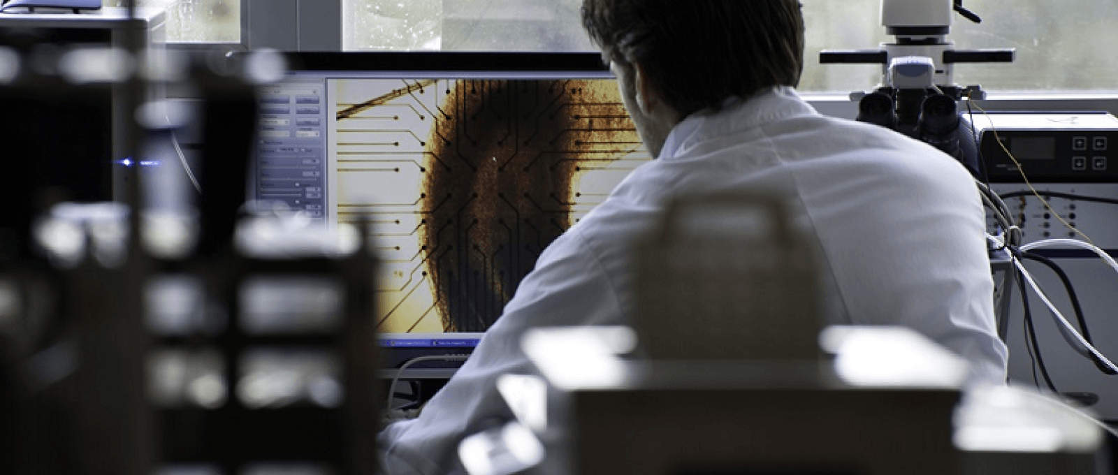
Pictures from the INMED laboratory


Research Teams
The INMED is composed by international researchers and professors brought together in 9 research teams.
A developmental scaffold for the organization of adult cortical networks
DescriptionMost adult cortical dynamics are dominated by a minority of highly active neurons distributed within a silent neuronal mass. If cortical spikes are sparse, spiking of single distinct neurons, such as hub neurons (Bonifazi et al. 2009), can impact on network dynamics and drive an animal’s behavior. It is thus essential to understand whether this active and powerful minority is predetermined and if true to uncover the rules by which it is set during development. Current work in the lab aims at testing the possibility that birthdate is a critical determinant of neuronal network function into adulthood, in health and disease.
More specifically, we reason that neurons that are born the earliest are primed to participate into adult network dynamics. This hypothesis is considerably fed by our past work aiming at understanding how cortical networks function and assemble during development. To test this hypothesis, and more generally to describe structure-function relationships in cortical networks, we have developed a multidisciplinary approach that combines in vitro and in vivo calcium imaging and electrophysiology, neuroanatomy, notably from clarified intact structures, mathematical analysis and mouse genetics.
Rosa Cossart Total : 2 HDRs
- Two-photon calcium imaging (in vitro and in vivo)
- Electrophysiology (in vitro and in vivo)
- Optogenetics
- Animal surgery, stereotaxy
- Electroencephalography (EEG)
- Computational Neuroscience
- Mouse genetics
- Neuroanatomy
- Light-Sheet Microscopy
- Developmental systems neuroscience
- Hippocampal circuit function
Development, Hippocampus, GABA, cortex, circuits, electrophysiology, memory, neuronal coding, epilepsy
Neuronal coding of space and memory
DescriptionThe hippocampus is important for our capacity to locate ourselves and navigate in familiar environments (spatial navigation). Our team is interested in understanding spatial navigation and its link with episodic memory formation. We address this question at the behavioral, network and cellular levels. At the behavioral level we train animals to navigate in real or virtual environments. Compared to real environments, virtual reality environments allow a better control of external sensory cues available to the animal to locate itself within the environment.
At the network level we use extracellular electrodes (e.g. silicon probes) to record the spiking activity of hundreds of cells in the hippocampal formation (hippocampus, entorhinal cortex) while animals are navigating. Because extracellular recordings can only record the spiking output of neurons but not the intracellular mechanisms leading to that spiking (synaptic inputs and intrinsic properties) we also recently contributed to the development of a new technique allowing to perform intracellular patch-clamp recordings of hippocampal neurons in navigating animals (Epsztein et al., 2011). Intracellular recordings allow us to study the cellular mechanisms of spatial coding in great detail.
- Immunostaining, histology, or flow cytometry
- Microscopy
- Electrophysiology (on slices or cells)
- Electrophysiology (in vivo)
- Animal surgery, stereotaxy
- Animal behavior
- Optogenetics
- Virtual reality
- Role of local sensory cues in setting hippocampal spatial coding resolution (extra VR)
- Role of self-motion cues and external visual cues in grid cells activation (extra VR)
- Role of intrinsic neuronal excitability in place cells’ activation (patch anesth/VR)
- Effect of locomotion on membrane potential dynamics of hippocampal principal cells (patch VR)
Network, hippocampus, behavior, virtual reality environment, spatial navigation, memory, episodic memory.
- Animal Cognition And Behavior
- Excitability, Synaptic Transmission, Network Functions
Early activity in the developing brain
DescriptionOur team is interested in the neuronal network activity expressed in the brain at the early developmental stages. In particular, we study the generation of the patterns of activity in the sensory (somatosensory and visual) cortices, with the aim to understand the neuronal network mechanisms of the earliest patterns, early gamma oscillations and spindle-burst, and their roles in the activity-dependent formation of the cortical maps. Consequently, we extrapolate our hypothesis made in the animal models to the human premature neonates, with the aim to understand how the brain operates during fetal stages. We are also studying the developmental changes in GABAergic neurotransmission, and its roles in the generation of physiological and pathological activities in the developing and mature brain after trauma (hypoxia, epilepsy and tramatic brain injury).
Par conséquent, nous extrapolons notre hypothèse faite dans les modèles animaux, aux nouveau-nés humains prématurés, dans le but de comprendre le fonctionnement du cerveau au cours des étapes du fœtus. Nous étudions également les changements développementaux dans la neurotransmission GABAergique et ses rôles dans la génération d’activités physiologiques et pathologiques dans le cerveau en développement (hypoxie, épilepsie et douleur).
Roustem Khazipov Total : 2 HDRs
- Molecular biology (PCR…)
- Biochemistry (Western blot…)
- Cell culture
- Immunostaining, histology, flow cytometry
- Microscopy (fluorescence, confocal, electronic…)
- Calcium imaging
- Electrophysiology (on slices or cells)
- Animal surgery, stereotaxy
- Pharmacology
- Animal behavior
- Movement or posture analysis, electromyography (EMG)
- Optogenetics
- Electroencephalography (EEG)
- Bioinformatics
- Physiological patterns of activity in the developing brain
- Developmental changes in GABA signaling
Epileptic activity in the developing brain - Neuroprotection of newborn during delivery
- Secondary neurogenesis in link with post traumatic depression
Development, neonate, electroencephalogram, cortex, barrel system, visual system, GABA, epilepsy, hypoxia, depression, secondary neurogenesis, oxytocin, fetal alcohol syndrome, chloride transporters.
- Animal Cognition And Behavior
- Computational Neuroscience
- Development Of The Nervous System
- Disorders Of The Nervous System
- Excitability, Synaptic Transmission, Network Functions
- Motor Systems
- Novel Methods And Technology Development
- Sensory Systems
Adolescence and developmental vulnerability to neuropsychiatric diseases
DescriptionOur general aim is to understand how meso-corticolimbic (MCL) microcircuits are shaped throughout early life critical periods especially adolescence, to give rise to harmonious emotional behaviors and cognitive functions in adulthood. Specifically, we want to understand how environmental and genetic insults modeling neuropsychiatric diseases transform the architecture and the functionality of synaptic networks and reduce the behavioral working range.
Our previous work fueled the concept that structural and functional damages during early life periods including adolescence are causal in disease-linked behavioral deficits. Our core hypothesis is that adolescence delineates a period of maximal vulnerability and consequently is a critical determinant of how environments and genes shape neuronal network functions into adulthood (Bara et al. 2018; Labouesse et. al. 2017; Manduca et al. 2017; Bouamrane et al. 2017; Iafrati et al. 2016; Iafrati et al. 2014).
Our research project will allow disambiguating complex phenotypes into new developmental endophenotypes and rational design of innovative therapeutic strategies
Olivier Manzoni, Pascale Chavis Total : 3 HDRs
- Microscopy
- Calcium imaging
- Electrophysiology
- Animal behavior
- Optogenetics
- Neurophysiology synaptic networks
- Neuropsychiatric diseases
Synapse, synaptic plasticity, accumbens, prefrontal cortex, endocannabinoid, mGluR, pharmacotherapy, addiction, autism, Fragile X, nutrition, adolescence, omega 3
- Animal Cognition And Behavior
- Development Of The Nervous System
- Disorders Of The Nervous System
- Excitability, Synaptic Transmission, Network Functions
The neural bases of sensorimotor learning
DescriptionHumans and animals’ survival depends on their ability to learn new adaptive behaviours, to adjust them to changes in their environment or occurring in their own body, and, in some cases, to become extremely skilled or efficient at what they do. All these functions depend on intricate interactions between sensory, motor, and more “cognitive” processes. The cortico-striatal system has been linked with learning but its exact contribution to the multiple processes occurring during different forms of learning and adaptation is far from being well understood.
Understanding the function(s) of the cortico-striatal system is important because its dysfunction results in several prevalent brain diseases such as Parkinson’s disease, mental retardation, or hyperactivity disorders that, noticeably, are characterized by a mixture of sensory, motor, and cognitive deficits.
Thus, our team tries to delineate the contribution(s) of the cortico-striatal system during learning and adaptation. We tackle this challenging objective by developing original tasks allowing us to capture and manipulate the behaviour of rodents. We combine this behavioural approach with a wide range of system neuroscience techniques (both in vivo during behaviour and ex-vivo) and computational/theoretical approaches.
David Robbe, Ingrid Bureau, Elodie Fino Total: 2 HDR
- Electrophysiology (in vivo, in vitro)
- Histology/Anatomy
- Animal surgery, (stereotaxic injection of excitotoxic compounds, virus, tracers)
- Animal behavior
- Movement or posture analysis (EMG)
- Optogenetics
- Electroencephalography (EEG)
- Data Analysis/Modeling
- Programming
Neuronal bases of sensorimotor learning and motor control.
Learning, basal ganglia, barrel cortex, motor cortex, neuronal coding, circuits, behavior, electrophysiology, optogenetics, multi-electrode.
- Animal Cognition And Behavior
- Disorders Of The Nervous System
- Excitability, Synaptic Transmission, Network Functions
- Motor Systems
- Sensory Systems
NICE2: Neonatal, Infantile and Childhood Epilepsies and Encephalopathies
DescriptionGenetic and nongenetic (viruses, drugs…) factors may cause or influence a broad range of neurodevelopmental disorders, including severe epilepsies and encephalopathies, which can be associated with comorbid manifestations (e.g. cognitive or behavioural impairment).
Despite the recent identification of various genes participating in such disorders, the underlying pathogenic mechanisms and rationale for treatment still remain poorly understood. In these conditions, identifying the early events likely altered during brain development and deciphering the underlying pathophysiological processes is mandatory.
The NICE2 team aims at studying and at targeting the early pathophysiological events associated with epilepsies and encephalopathies of genetic or nongenetic origin, using multidisciplinary approaches in vitro and in vivo.
We are particularly interested in the pathological alterations that occur early in the developing brain and that may have profound and long-term consequences on brain development and functioning.
Our main objectives are:
- to better understand pediatric epilepsies and epileptic encephalopathies of genetic origin, and to design novel rescue strategies in those contexts;
- to investigate secondary pathophysiological events, such as neuroimmune alterations (e.g. microglia dysfunctioning), that are likely to impact on the severity, the comorbidity, the outcome and the responses to treatments;
- to decipher how nongenetic factors (e.g. viral infections) impact on brain development and ultimately lead to neurodevelopmental disorders.
Pierre Szepetowski Total : 3 HDRs.
- Molecular biology
- Biochemistry
- Cell culture
- Immunostaining, histology, flow cytometry
- Microscopy (confocal, 2-photon)
- Electrophysiology (in vitro, in vivo)
- Animal surgery, stereotaxy
- In utero electroporation
- Animal behavior
We study four rodent models of different neurodevelopmental disorders where both specific and non-specific determinants are likely involved, albeit at different levels, in the emergence and in the variable evolution of the phenotypes. Those disorders are:
- KCNQ2-related early-onset epileptic encephalopathies
- GRIN2A-related disorders
- TSC1-related disorders
- Cytomegalovirus (CMV)-related disorders
Epilepsy / Encephalopathies /Brain development / NMDA Receptors / GRIN2A / Kv7.2 / Tuberous sclerosis complex /Congenital cytomegalovirus / Microglia / Rodent models
- Animal Cognition And Behavior
- Development Of The Nervous System
- Disorders Of The Nervous System
Neuronal coding and plasticity in epilepsy
DescriptionOur team is interested in the coding of information in the hippocampus, a structure involved in memory. We focus our works on the dentate gyrus, sitting between the entorhinal cortex and area CA3. This region is both anatomically well positioned and physiologically predisposed to play the role of a gate, blocking or filtering excitatory activity from the entorhinal cortex. We investigate the neuronal computation and plasticity of dentate granule cells in normal and pathological conditions. Our studies are conducted at multiscale levels i.e. from the individual spine to the microcircuit. We also analyze the role of cell adhesion molecules implicated in ion channel positioning which are implicated in autoimmune diseases.
Valérie Crépel Total : 2 HDRs
- Electrophysiology
- Cell culture
- Calcium imaging
- Immuno-histochemistry
- Animal behavior
- The coding of information in the hippocampus
- The neuronal computation and plasticity of dentate granule cells in normal and pathological conditions
- The role of cell adhesion molecules
hippocampus, rodent, lobe epilepsy, autoimmune diseases, dentate gyrus.
- Animal Cognition And Behavior
- Disorders Of The Nervous System
- Excitability, Synaptic Transmission, Network Functions
Molecular basis and pathophysiology of cortical development disorders
DescriptionOur team investigates Malformations of Cortical Development (MCDs) which are important causes of mental retardation and account for 20-40% of drug-resistant childhood epilepsy.
We conduct integrated multidisciplinary studies involving morphologists, molecular biologists and electrophysiologists. In addition, we have established collaborations with clinicians and geneticists (European EPICURE project) providing us with a transversal appreciation of our researches.
We concentrate our efforts on:
- the identification of new genes and molecular actors involved in normal migration processes and being altered on MCDs;
- a better understanding of the link between genotype and phenotype, and of the process leading to an abnormal migration pattern;
- the characterization of the physiopathological mechanisms responsible for epileptogenesis in MCDs, in order to precisely identify the seizure-generating zone, to describe its properties and the mechanisms of seizure generation, and to ultimately suggest new therapeutic approaches.
- Histological and Neuroanatomical Methods,
- Cellular and Molecular Biology (RNAi interference, in utero electroporation),
- Cell and Slice Culture,
- Extracellular Multi-site Recordings with
- Microelectrode Arrays,
- Time-lapse Imaging
- Identification of new candidate genes for neuronal migration and MCDs
- Mecanisms of genesis of MCDs
- Morphofunctional analysis of heterotopic networks
- Functional repercussions of cortical migration disorders: analysis of DCX knockdown rats
Brain development, Epilepsy, Cerebral Cortex, Malformation of Cortical Development, Neuronal Migration, Animal Models.
Early life imprinting and neurodevelopmental disorders
DescriptionPrader-Willi Syndrome (PWS) is a complex and rare neurogenetic disease characterized by a range of physiological, endocrine, cognitive and behavioural disturbances, which might be considered as a lack of adaptation of the organism to the environment. The cause of this disease is genetic and the candidate genes are all regulated by the genomic imprinting, a mechanism leading to a paternal expression of those genes, the maternal alleles being normally silenced. In our team, using mouse models genetically modified, we study the physiopathological and physiological role of two candidate genes involved in PWS: the Necdin and Magel2 genes; both genes belong to the same family of MAGE genes whose the function is still enigmatic.
Our project focused on the phenotype investigation of these mouse mutants. The abrogation of Necdin leads to a partial early post-natal lethality due to respiratory distress (in 30% of cases), to a growth retardation, sensory-motor defects and behavioural alterations. Cellular investigations of the sensory and motor deficits revealed that Necdin is an anti-apoptotic factor in developing sensory neurons and motor neurons. On the other hand, investigations of the breathing deficit in Necdin KO mice showed an alteration of the regulatory systems of the Respiratory Rhythm Generator (RRG); a dysfunction of the serotonergic system in Necdin-KO mice, known to play a crucial role in maturation and function of the central respiratory system, might be the cause of this deficit Magel2 paternal deletion results in a fatal neonatal failure to thrive due to feeding problems; this phenotype is rescued by oxytocin administration.
Our results show a crucial role of oxytocin in feeding behaviour at birth. Adult mutant mice bypassing this lethality display growth retardation, hormonal, metabolic and circadian activity alterations.These in vivo studies on mouse models are very important to understand the PWS. Beyond that, they reveal new physiological, cellular and molecular neuronal pathways involve in vital functions, which might be altered in other diseases.
- Creation and studies of mouse models genetically modified.
- Histology,
- Cellular and molecular biology,
- Imaging,
- Stereotaxia,
- Virus injection.
- Oxytocinergic Alterations
- Serotonopathy
- Genetics and Epigenetics
Genomic imprinting, Prader-Willi, Necdin, Magel2, oxytocin, feeding behaviour, serotonin, respiratory distress.


