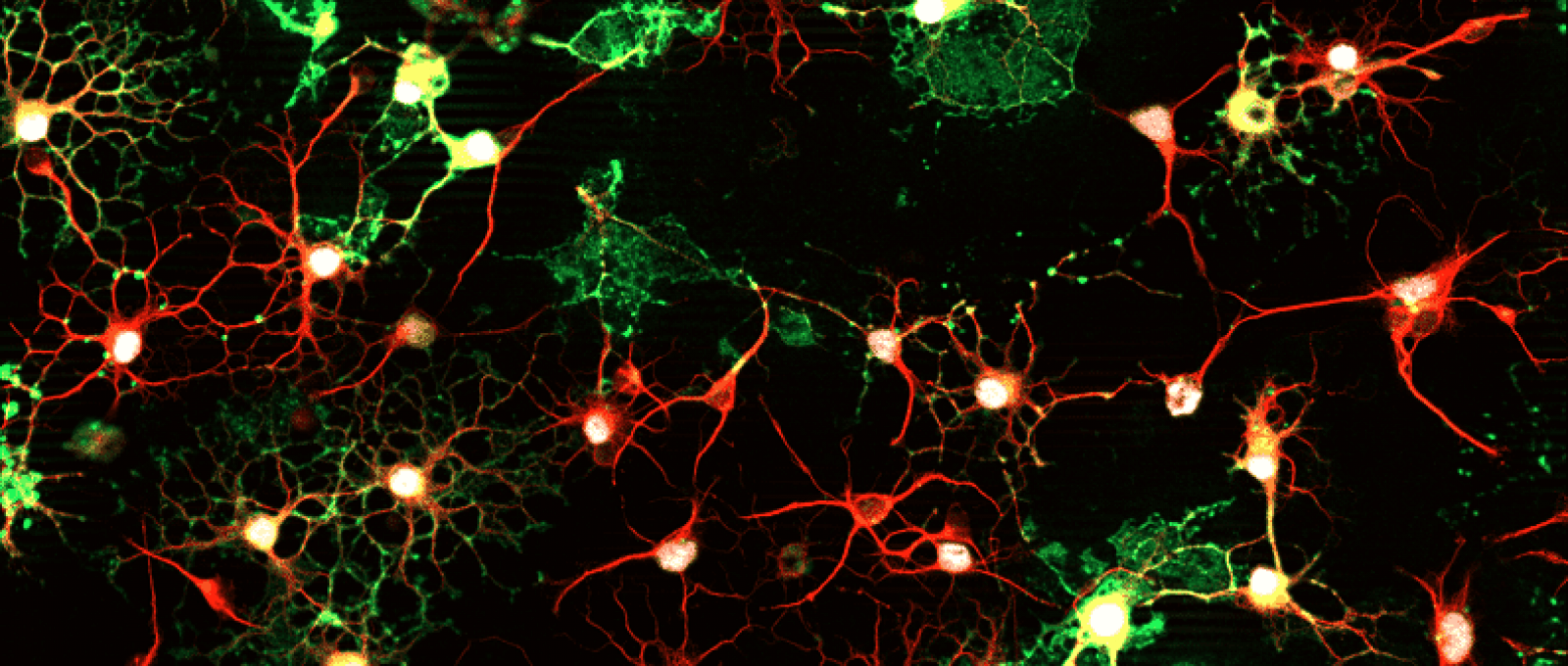
Oligodendrocytes en culture : myéline (protéine MBP, en vert), cytosquelette (tubuline, en rouge) et noyaux (Olig2, en blanc). © AMU/CNRS Karine Magalon (IBDM)

Developmental Biology Institute of Marseille

IBDM is an interdisciplinary research institute oriented towards developmental biology and pathologies.

Half of its twenty teams addresses biological questions related to the nervous system development, maturation and plasticity. Unraveling these processes is essential to understand the pathogenesis of neurological disorders and design novel therapeutic targets.

The institute encompasses complementary scientific approaches including experimental embryology, physiology, cellular/molecular biology, genetics, biocomputing and genomics.

Using a large spectrum of animal models, IBDM research effort in Neurosciences focuses on neural stem cell biology, cell fate determination, cell division and migration, axon guidance and circuit formation, neuroplasticity in the normal adult brain and adaptive changes in disease states.

The scientific equipment is grouped into innovative and effective technical platforms, including state of the art imaging center, animal housing and functional exploration facilities. One of the key objectives of the IBDM is to encourage interaction with different disciplines –mathematics, physics, chemistry – in order to develop new experimental approaches.

In addition to basic research, IBDM promotes projects in the field of applied science, aimed at developing therapeutic molecules. Furthermore, in collaboration with the university, the institute plays an important role in developing teaching programmes in biology and at the interface between biology and other disciplines.

Pictures from the IBDM laboratory

Research Teams

Discover below the 11 IBDM teams working on neuroscience issues ranging from neural stem cell biology to function and plasticity of neural networks to understand the interplay between development, physiology and pathologies of the nervous system.

Visit the IBDM's electron microscopy platform in this 360° video (don't forget to enable the English subtitles).
21Teams 50Members10000Publications

Stem cells and brain repair

Description

The team focuses on the process of post-lesional plasticity following demyelination in the adult brain. In some patients affected by multiple sclerosis (MS) spontaneous remyelination can occur. Although insufficient to counter the damages caused by the repetitive attacks, this spontaneous regenerative process represent great therapeutic hopes. Studies led on rodent have identified two distinct sources of cells involved in remyelination: the oligodendrocyte progenitors (OPCs) found throughout the brain parenchyma and neural progenitor derived from the sub-ventricular zone (SVZ) were adult neural stem cells reside.

Our objectives are to uncover the cellular and molecular mechanisms controlling this process in order to increase our fundamental knowledge on brain regeneration and to promote myelin repair.

Team leader

Pascale Durbec  Total : 2 HDRs.

Techniques

  • Molecular biology
  • Biochemistry
  • Cell culture
  • Immunostaining, histology, or flow cytometry
  • Microscopy
  • Animal surgery, stereotaxy
  • Animal behavior

Research axes

  1. The cellular and molecular control of myelin formation and regeneration in the adult rodent brain.
  2. The contribution of adult stem cells to brain repair.
  3. The mechanisms that regulate oligodendrocytes migration and differentiation.
  4. The research and preclinical evaluation of new therapeutic strategies for myelin repair in patients suffering from multiple sclerosis.

Keywords

Adult stem cell, migration, myelin, neurodegeneration, oligodendrocyte, plasticity, remyelination, multiple sclerosis, experimental models, experimental therapeutics

Thematics
  • Animal Cognition And Behavior
  • Development Of The Nervous System 
  • Disorders Of The Nervous System

Cellular interactions, neurodegeneration and neuroplasticity

Description

The team studies cellular interactions and neuroplasticity in the adult brain, in particular in the context of basal ganglia-related functions and pathologies.

Ongoing projects are centered on the pathophysiology and treatment of Parkinson’s disease and the role of glutamatergic systems.

Our approach combines the use or development of relevant experimental models in rodents (chronic deep brain stimulation, genetic model of cell specific ablation, optogenetic modulation of neuronal activity) with a variety of analytic tools including functional anatomy, electrophysiology (in vitro and in vivo), neurochemistry and molecular/cellular biology.

Team leader

Lydia Kerkerian-Le Goff Total : 5 HDRs.

Techniques

  • Molecular biology
  • Biochemistry
  • Immunostaining, histology, or flow cytometry
  • Microscopy
  • Electrophysiology (on slices or cells)
  • Electrophysiology (in vivo)
  • Animal surgery, stereotaxy
  • Pharmacology
  • Animal behavior
  • Optogenetics
  • Deep brain stimulation

Research axes

  1. The anatomofunctional organization of the basal ganglia network and its remodeling in Parkinson’s disease
  2. The cellular/molecular bases of the beneficial or side-effects of the current treatments for Parkinson’s disease
  3. The research and preclinical evaluation of new therapeutic strategies
  4. The cell death mechanisms, with focus on those linked to dysfunction of glutamate transporters

Keywords

Neuroplasticity, basal ganglia, Parkinson’s disease, experimental models, experimental therapeutics, dopamine, glutamate, excitotoxicity, neurodegeneration, physiopathology

Thematics
  • Animal Cognition And Behavior
  • Excitability, Synaptic Transmission, Network Functions
  • Motor Systems
  • Novel Methods And Technology Development

Chronic pain: molecular and cellular mechansims

Description

Our team aims at understanding how primary sensory neurons diversity is translated at the functional level. To do this we use state-of-the-art technologies combining mouse genetics, transcriptomic analyses, molecular and cellular biology, electrophysiology and behavior.  In the last few years we successfully contributed to extend our understanding of the molecular logic that governs nociceptive neurons diversity, we unraveled the functional role of newly identified genes in pain biology and deciphered the functional specialization of two newly characterized subpopulations of primary sensory neurons. Currently, our projects are heading towards a better understanding of the circuitry between primary sensory neurons and spinal cord neuronal networks. We are also developing clinically-driven projects in which we investigate the molecular and cellular mechanisms that underlie the transition from acute to chronic pain.

Team leader

Aziz Moqrich  Total : 2 HDRs.

Techniques

  • Molecular biology
  • Biochemistry
  • Cell culture
  • Immunostaining, histology, or flow cytometry
  • Microscopy
  • Calcium imaging
  • Electrophysiology (on slices or cells)
  • Animal surgery, stereotaxy
  • Pharmacology
  • Animal behavior
  • Optogenetics
  • Bioinformatics
  • Mouse genetics

Research axes

  1. Primary sensory neurons diversity
  2. Role of DRG neurons subset in pain sensation
  3. Dicephiring the circuitry between C-LTMRs and laminae IIi interneurons
  4. Molecualr and cellular mechanisms underlying the transition from acute to chronic pain

Keywords

Primary sensory neurons, neuronal circuits, behavior, pain, disease, neurogenesis, transgenic mice.

Thematics
  • Animal Cognition And Behavior
  • Development Of The Nervous System 
  • Excitability, Synaptic Transmission, Network Functions

Polarization and binary cell fate decisions in the nervous system

Description

In both vertebrates and invertebrates, postmitotic neurons are often generated by asymmetric divisions of neuronal progenitors such as neural stem cells. This general mechanism used to build the nervous system raises two important questions : how are these asymmetric divisions coordinated in space and how do the daughter cells acquire different fates.

We address these questions using the nematode C. elegans as a model organism. In C. elegans, most neurons are generated during neurulation by asymmetric divisions oriented along the antero-posterior axis. We recently showed that these terminal asymmetric divisions are regulated by a particular Wnt/β-catenin pathway. We are now trying to understand :

1) How the field of neuronal precursors is polarized.

2) How the daughter cells acquire different fates and especially how the asymmetric division machinery is connected to the terminal differentiation program of postmitotic neurons.

The Wnt/β-catenin pathway is involved in several types of cancer and in the regulation of asymmetric divisions of neural stem cells in vertebrates. This study may therefore help identify candidate target proteins and mechanisms for future anti-cancer drug developments or regenerative medicine treatments.

Team leader

Vincent Bertrand Total : 1 HDR.

Techniques
  • Molecular biology
  • Biochemistry
  • Cell culture
  • Immunostaining, histology, or flow cytometry
  • Microscopy

This team is not affiliated to the Neuroscience Master’s.

Research axes

  1. Polarization mechanism of a neuronal precursor field
  2. Role of the Wnt pathway in the specification of neuronal subtype identities
  3. Role of chromatin factors in neuronal differentiation
  4. Evolution of the mechanisms of neuronal specification

Keywords

Advanced in vivo imaging, asymmetric division, Caenorhabditis elegans, gene regulation, neurogenesis, neuronal precursors, stem cell, tissue polarity, Wnt signaling

Thematics
  • Development Of The Nervous System 
  • Disorders Of The Nervous System

Molecular control of neurogenesis

Description

How are neural stem cells determined to generate neurons with defined neurotransmitter phenotypes? How do neurons migrate to their final positions? How are neurons synaptically integrated into the circuitry and what is their physiological function?

We use postnatal olfactory bulb neurogenesis to identify and functionally characterize factors and signaling cascades that regulate these processes. To reach this aim we combine high-resolution gene expression screens with mouse genetics and state-of-the-art in vivo brain imaging by multiphoton microscopy.

Team leader

Harold Cremer  Total : 4 HDRs.

Techniques

  • Molecular biology
  • Biochemistry
  • Cell culture
  • Immunostaining, histology, flow cytometry
  • Microscopy
  • Calcium imaging
  • Animal surgery, stereotaxy
  • Brain imaging – Animal
  • Bioinformatics

Research axes

  • Neural stem cells

Keywords

Adult neurogenesis, cell therapy, dopaminergic neurons, in vivo electroporation, microRNAs, neurogenesis, Parkinson’s disease, stem cell, transgenic mice

Thematics
  • Development Of The Nervous System 
  • Excitability, Synaptic Transmission, Network Functions
  • Sensory Systems

Transcriptional regulatory network in development and diseases

Description

Teashirt3 (TSHZ3) is a zinc finger transcription factor whose targets and functional implications remain largely unknown. Our previous studies support that Tshz3 plays a role in the developing and maturing cerebral cortex. To unravel the role of Tshz3 in the development and function of cortical projection systems, we are characterizing novel mouse models of Tshz3 deletion by combining mouse genetics, molecular biology, morphological and tract tracing studies, slice electrophysiology and behavioral approaches. This research will improve our knowledge on mechanisms underlying early corticogenesis and/or maturation and functioning of cortical circuits.

Team leader

Laurent Fasano  Total : 3 HDRs.

Techniques

  • Molecular biology, in situ hybridization
  • Biochemistry
  • Cell culture
  • Immunostaining, histology
  • Microscopy
  • Animal behavior
  • Bioinformatics

Keywords

Behavior, transcriptional control, corticogenesis, development, differentiation, neuronal identity, disease, morphogenesis, visceral smooth muscle, cortical neurons, gene regulation

Thematics
  • Animal Cognition And Behavior
  • Development Of The Nervous System 
  • Disorders Of The Nervous System

Development and pathologies of neuromuscular circuits

Description

The Helmbacher team studies neuromuscular development and pathology. Our research aims to understand processes that control the development of neuromuscular circuits, and to uncover how alterations of these developmental processes lead to devastating neuromuscular pathologies in human. Our work on neuromuscular development integrates the study of cell fate diversity and the mutual dependency of motor neurons and their target muscles. We aim to identify the signals successively exchanged by neurons and muscles during development, such as the signals acting on neuronal and muscular specification, the signals that pattern neural projections through axonal guidance and through muscle morphogenesis, the signals that regulate homogeneous growth and match the size of neuromuscular units. We study how all these signals successfully integrate to produce locomotor activity.

Our work recently uncovered the link between a human myopathy, Facioscapulohumeral dystrophy, and genetic alterations of the FAT1 cadherin gene, a gene involved in neuromuscular development. We currently work at determining the contribution of FAT1-dependent phenotypes to FSHD-like muscular symptoms. Besides, we also explore how FAT/Dachsous cadherin signaling contributes to the functional maturation of neuromuscular circuitry.

Team leader Techniques

  • Molecular biology
  • Cell culture
  • Immunostaining, histology, or flow cytometry
  • Microscopy
  • Movement or posture analysis, electromyography (EMG)
  • Bioinformatics
  • Genetics (mouse models)

Keywords

  1. Mechanisms of neuromuscular circuit assembly
  2. Neuromuscular Fatopathies and FSHD (facioscapulohumeral muscular dystrophy) physiopathology
  3. Role of FAT/Dachsous signaling during functional maturation of neuromuscular circuits.

Keywords

Neural circuits, connectivity, neuromuscular development, muscular dystrophy, axon guidance, cell migration, adhesion molecule, morphogenesis, motor neuron, muscle, pathophysiology.

Thematics
  • Development Of The Nervous System 
  • Disorders Of The Nervous System
  • Motor Systems

Signalling networks for stemness and tumorigenesis

Description

We aim at uncovering how convergent instructive signals cooperate to regulate the fate of cells. This question is addressed by using two biological contexts: the transition of healthy cells towards tumorigenesis and the regulation of self-renewal versus differentiation of stem cells.

Concerning stem cells, the team studies how human induced pluripotent stem cells (iPSCs) fine-tune perception of instructive extracellular signals in order to: 1) enhance generation of functional neurons (e.g. dopaminergic neurons) both in vitro and in vivo; 2) improve safety of human iPSC-derived cell grafts in animal models of neurodegenerative disorders (e.g. Parkinson’s disease) by preventing tumour side effects.

Our objective is to acquire knowledge on human iPSC biology related to neural/neuronal differentiation by exploring molecular and signalling networks. This research may open new perspectives in regenerative medicine by developing more effective, user-friendly, and safe stem cell-based therapeutic strategies (project leader Dr Rosanna Dono).

Team leader

Flavio Maina  Total : 3 HDRs.

Techniques
  • Molecular biology
  • Biochemistry
  • Cell culture and embryo culture
  • Immunostaining, histology, or flow cytometry
  • Microscopy
  • Animal behavior
  • Bioinformatics
  • Transgenesis
  • Xenografts

This team is not affiliated to the Neuroscience Master’s.

Research axes

  1. Modelling receptor tyrosine kinase signalling in vivo to uncover cooperative signals in cancer.
  2. Balancing stem cells self-renewal versus differentiation by modulating perception of instructive signals.
  3. Signalling network crosstalk uncoupling tumorigenicity from therapeutic properties of human iPSCs.
  4. Exploring mechanisms underlying neuronal differentiation (e.g. dopaminergic fate acquisition) during mouse embyogenesis and in human iPSCs.
  5. Preclinical evaluation of human iPSCs-based therapy for neurodegenerative disorders, using Parkinson as disease model.

Keywords

Modelling tumorigenesis, receptor tyrosine kinase signalling in vivo, signalling modulation, glypicans, stem cells, human iPSCs, development, mouse genetics, neuronal differentiation, Parkinson’s disease, stem-cell based replacement therapy.

Thematics
  • Development Of The Nervous System 
  • Disorders Of The Nervous System

Axon plasticity in development and cancer

Description

Normal brain function depends on complex patterns of neuronal circuits that develop during fetal life and childhood. Neurons connect to each other by extending long cables, called axons, whose growth is not random but precisely oriented towards their targets by axon guidance molecules. Our team studies the mechanisms that contribute to the fine regulation of guidance cue activities to ensure the accuracy and fidelity of axonal trajectories.

In addition to being essential to brain development, axon guidance molecules are also present in adult organisms where their expression can be reactivated under pathological conditions such as cancer. We are investigating whether their activity could contribute to the innervation of malignant tumors, a process that is still poorly characterized but can influence the course of the disease.

Team leader

Fanny Mann  Total : 2 HDRs.

Techniques

  • Molecular biology
  • Biochemistry
  • Cell culture
  • Immunostaining, histology, flow cytometry
  • Microscopy
  • Animal surgery, stereotaxy
  • Brain imaging – Animal

Research axes

  1. the cellular and molecular events controlling the development of axonal projections
  2. the remodeling of axonal projections, their function and implication in cancers

Keywords

Neurodevelopment, neuronal circuits, axon guidance, intracellular trafficking, signaling, neurodevelopmental disorders, cancer, tumor axonogenesis.

Thematics
  • Animal Cognition And Behavior
  • Development Of The Nervous System 
  • Disorders Of The Nervous System

Neural stem cell plasticity

Description

Neural stem cells found in the nervous system are very plastic. This plasticity allows them to generate a vast repertoire of neural and glial progeny while the brain is being built or to regenerate neurons after brain injury. Our team aims at deciphering the molecular and genetic mechanisms underlying this plasticity. We are also investigating how environmental conditions during fetal growth affect neural stem cell plasticity and their ability to constitute their full repertoire of neurons. Finally, because of their plasticity and large proliferation potential, stem cells are largely exposed to cancer-promoting defects. Our ambition therefore consists in uncovering how neural stem cell plasticity can be hijacked for the benefit of cancerous processes. Understanding these basic principles could help correcting cellular failures responsible for cancer induction, delaying ageing and exploiting neural stem cell regenerative potential.

In order to investigate these fascinating issues, we use the fruitfly Drosophila. As in mammals, the Drosophila adult brain is mainly composed of neurons and glial cells (>100,000) disposed in complex neural circuits. These cells have been generated in the developing animal from a limited set of neural stem cells. We take advantage of the powerful genetic tools developed in this model organism to manipulate neural stem cells while the brain is being constructed during development. We aim at identifying the genes and molecular mechanisms controlling their properties.

Team leader

Cédric Maurange  Total : 1 HDR.

Techniques
  • Molecular biology
    Biochemistry
    Immunostaining, histology, or flow cytometry
    Microscopy
    Bioinformatics


This team is not affiliated to the Neuroscience Master’s.

Research axes

  1. to decipher how a genetic program is deployed in every neural stem cells to ensure that different types of neurons are generated over time
  2. to investigate the impact of nutritional conditions on the making of the brain
  3. to explore the mechanisms that drive tumour progression in a developing brain, as happens for pediatric neural cancers

Keywords

Cancer, neural stem cell, cancer stem cell, neuronal specification, brain development, nutrition, paediatric cancer, Drosophila

Thematics
  • Development Of The Nervous System 
  • Disorders Of The Nervous System

Effects of gut microbiota on host behavior

Description

The intestinal microbiota is composed of bacteria whose nature depends on the intestinal environment and the food preference of the host. In return, the microbiota manipulates the host by producing nutrients and excreting metabolites that modulate its physiology. If it is established that the microbiota regulates basal metabolism or educates the host’s immune system, recent results attest to its influence on the nervous system. Anxiety, depression, autism,… the microbiota modulates our behavior, regulates our emotions and intervenes in certain pathologies of the nervous system. If some of the mechanisms of host-microbiota communication have been elucidated, the field of investigation remains vast.

Thanks to the power of its genetics and the relative simplicity of its microbiota, Drosophila represents a promising alternative to mammalian models. Our results show that a bacterial component called peptidoglycan (PGN) is a key mediator in this dialogue. Under certain circumstances, PGN can cross the intestinal epithelium and reach the circulating blood in which organs and tissues are bathed. Our work has shown that the detection of this microbiota derived-PGN modulates the activity of certain octopaminergic neurons and ultimately the behavior of the host. Our team seeks to dissect the mechanistic details of this molecular dialogue between the microbiota and the host nervous system.

Team leader

Julien ROYET  Total : 2 HDRs.

Techniques

  • Molecular biology
  • Biochemistry
  • Cell culture
  • Immunostaining, histology, flow cytometry
  • Microscopy
  • Animal behavior

Research axes

  1. Understanding how PGN reaches and is sensed by octopaminergic neurons
  2. Understanding how the perception of PGN by these neurons changes their activity
  3. Understanding why only few host neurons respond to microbiota derived PGN

Keywords

Behavior, drosophila, octopaminergic neurons, microbiota, peptidoglycan

Thematics
  • Animal Cognition And Behavior
  • Motor Systems
  • Sensory Systems

Evolution and Development of morphology and behavior

Description

Team leader

Benjamin Prudhomme  Total : X HDRs.

Techniques

  • behavioral assays,
  • genetics & neuronal imaging,
  • calcium imaging,
  • functional transgenesis in various Drosophila species.

Research axes
Keywords
See more
Your opinion is important to us

Description de la soumission d'un avis
Your vote :
Your opinion is important to us
Publications